Vertebral compression fractures can be severely painful and life disrupting. Frequently this pain causes patients to go to bedrest which can lead to life threatening complications, such as blood clots, pneumonia, and structural changes restricting breathing or balance.
Fortunately, there are minimally invasive interventions that can stop the pain so that out of bed activity can be restored.
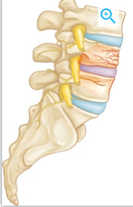
drawing of a vertebral body partial collapse due to fracture
The vertebral body has a significant nerve supply. When its fractured, severe pain can occur. This is increased when pressure from being upright (standing, walking, twisting, bending) is happening. This is no different than walking around on a broken leg or hip. Stabilizing the fracture stops the pain.
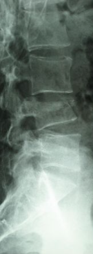
Lateral x-ray of lumbar vertebral body fracture.
Most vertebral compression fractures can be stabilized in a minimally invasive way. Using a fluoroscope (x-ray) bone cement can be injected into the marrow cavity of the vertebral body. This can be done under local anesthesia with or without a light sedation.
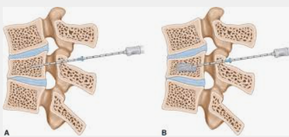
Drawing of needle placement into the vertebral body (A) with bone cement (B)
The bone cement injected into the vertebral body stabilizes the fracture curing approximately ten minutes after its administration (Vertebroplasty). If the fracture is diagnosed early, it may be possible to restore the square shape of the vertebrae in addition to stabilizing the fracture (Kyphoplasty).
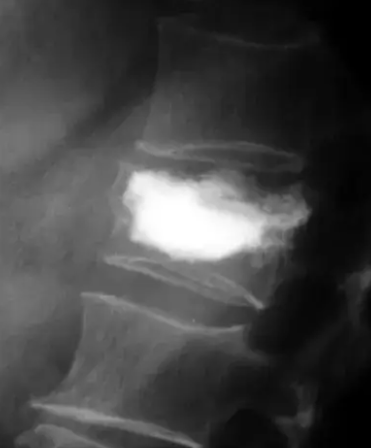
Bone cement injected after a balloon re-established the square shape of the vertebral body.
This minimally invasive procedure is performed as an outpatient. The cement completely cures in approximately ten minutes, with the patient in the recovery room for one hour before walking and discharge.